Mammogram
What is a mammogram?
A mammogram is an X-ray picture of the breast. There are two main types of mammograms: screening mammograms and diagnostic mammograms. A screening mammogram is done to check for breast cancer in a woman who has no signs or symptoms of the disease. If a woman has a lump or another sign or symptom of breast cancer (such as nipple discharge, thickening of the skin of the breast, pain in the breast or a change in the breast’s size or shape), the exam is called a diagnostic mammogram. A diagnostic mammogram may also be scheduled to evaluate changes seen on a screening mammogram.
All Northwell Health breast imaging centers offer 3D mammography, also known as tomosynthesis. This advanced form of mammography uses a low-dose X-ray system and computer reconstruction to create three-dimensional images of the breast. For a conventional mammogram, only two views are taken: from top to bottom, and from side to side which causes overlapping of the breast tissue. With 3D mammography, multiple images are taken of each breast from many different angles, and less pressure is applied—just enough to keep the breast in position. The multiple images are then assembled by a computer to produce clear images of the tissue throughout the breast. Because 3D mammography enables the examination of breast tissue one layer at a time, there is less chance that an abnormality will be obscured or overlooked. It also reduces the risk that overlapping tissue will create the appearance of an abnormality that isn’t really there—a false alarm.
Northwell Health breast imaging centers use the most advanced reconstruction software to interpret our 3D mammograms. This software allows us to perform 3D mammography with no more radiation than you would receive from a traditional mammogram.
Why it's done
Screening mammography aids in the early detection of breast cancer, when in many cases it is more curable and when breast-conserving therapies are available. Studies show that it can reduce the risk of death from breast cancer for women ages 40 to 74. Northwell Health Cancer Institute recommends that women be screened with mammography annually, starting at age 40.
Diagnostic mammography is used to evaluate a patient with abnormal clinical findings, such as a breast lump or nipple discharge. It may also be done if a screening mammogram identifies an abnormality or other area of concern.


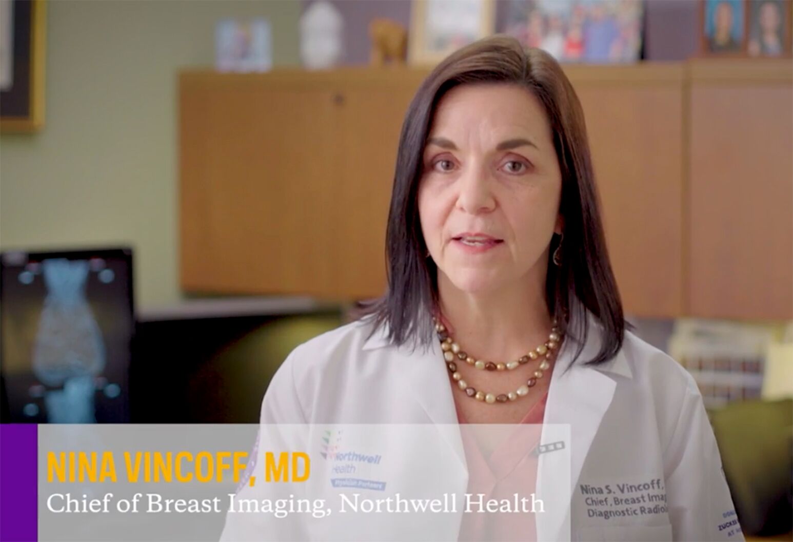
Women at average risk should start annual mammograms at age 40. Dr. Nina Vincoff shares four tips.
Over the course of a lifetime, one in eight women will be diagnosed with breast cancer. Take the breast cancer risk assessment to determine your risk.

View our educational brochure to learn more about breast density and why it matters.
Learn about our mammogram and other imaging services.
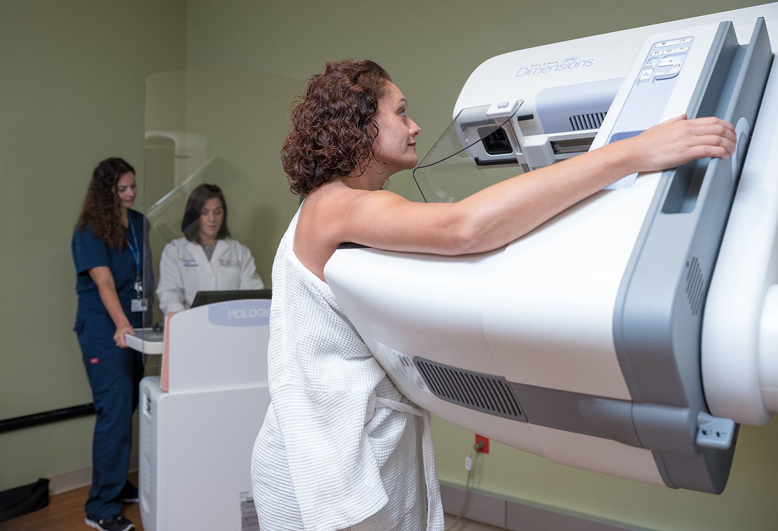
Our approach
When it comes to breast mammography, the breast radiologists at Northwell Health Imaging are the power behind the exam. Every mammogram is performed with state-of-the-art technology and interpreted by a fellowship-trained breast radiologist whose diagnostic skills have been honed by specialized training and thousands of hours of experience. Our nurse navigators serve as resources, providing support while guiding you through the process of assuring the best possible breast health, and our certified mammography technologists are highly skilled and experienced in positioning to put you at ease and to help you feel as comfortable as possible throughout the exam. If you need additional care, your breast radiologist will work side by side with the rest of your breast care team, collaborating closely to answer your questions and help guide your care.
Northwell Health Imaging offers the largest group of fellowship-trained and subspecialized breast radiologists on Long Island, as well as access to all the resources and clinical expertise of New York state’s largest health system. Whether you are here for screening, diagnostic or treatment imaging services, each of our practitioners is committed to providing a caring, comfortable environment and a positive, productive experience.
Risk factors
Excessive exposure to radiation raises the risk of cancer. However, the potential benefits of screening or diagnostic mammography outweigh the potential risks.
Studies show that 5 to 15 percent of screening mammograms require follow-up testing, such as additional mammograms or ultrasound, and in some cases, those tests prompt a biopsy. It is important to know that if your doctor recommends a biopsy, it does not necessarily mean you have cancer. In fact, most of these biopsies show that no cancer is present.
If there is a chance you might be pregnant, speak to your doctor before having a mammogram. While it is thought to be safe to have a mammogram if necessary during pregnancy, your doctor may want to arrange for extra protection, such as a lead shield over the lower part of the belly, or may recommend a different test.
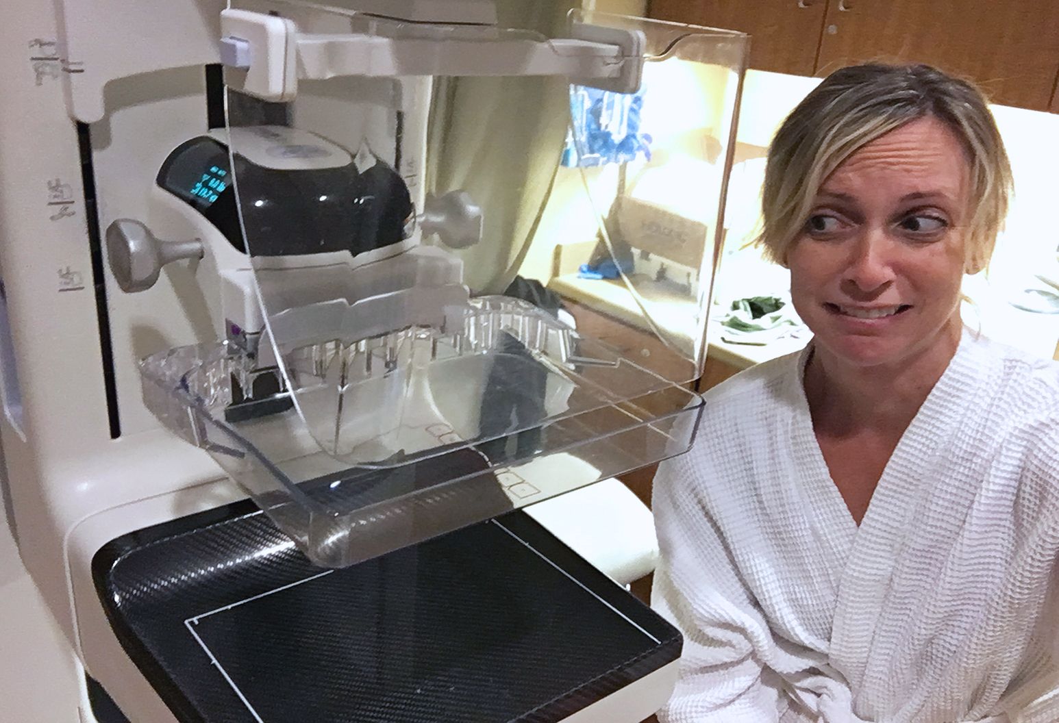
What to expect
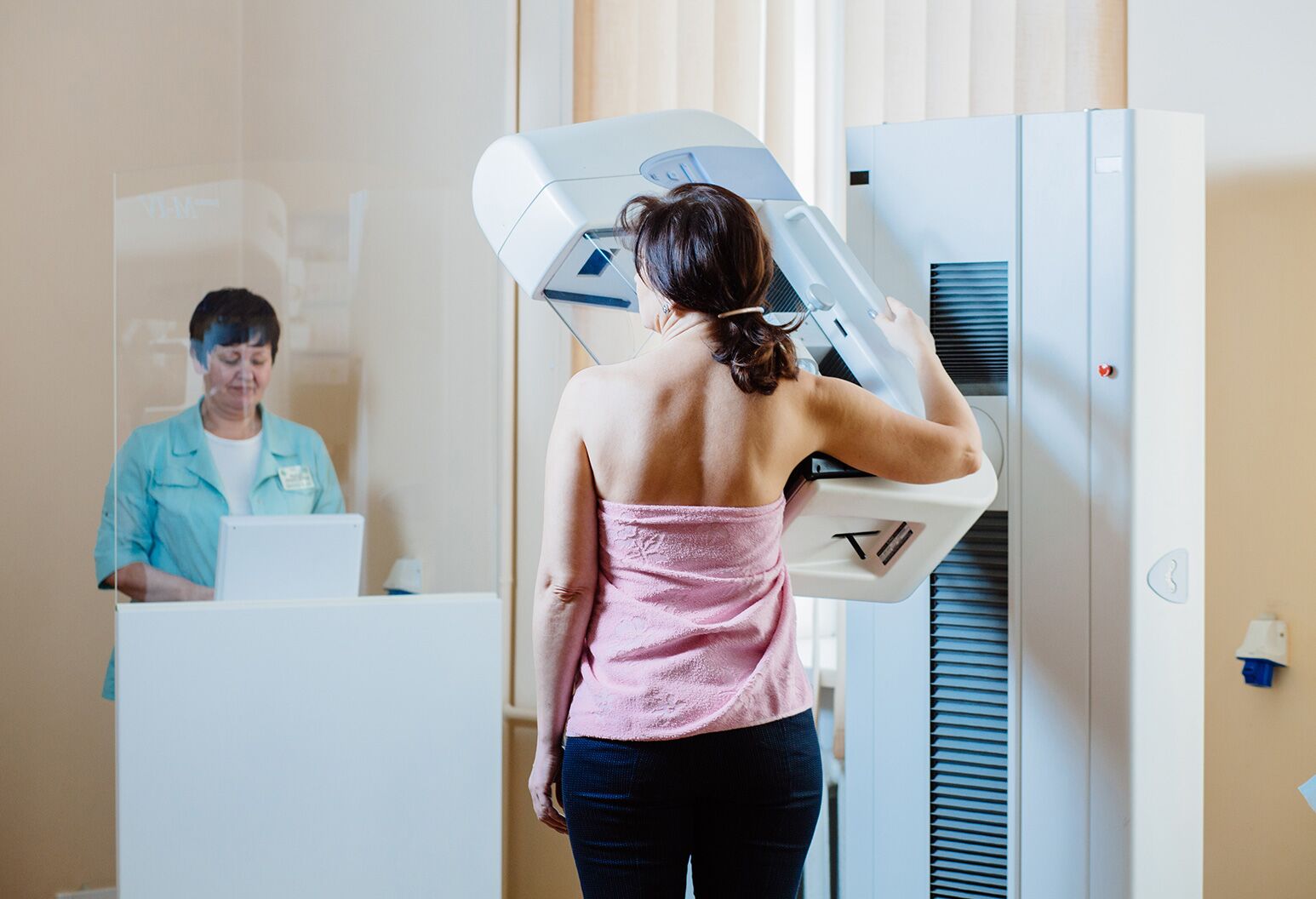
Mammography is performed on an outpatient basis. A technologist will position and compress your breast on a special platform. Compression is necessary for both 2D and 3D mammography. Among other things, compression reduces the chance that potential abnormalities will be hidden by overlapping breast tissue, and reduces movement of the breast, which can blur the image.
You will be asked to change position between images. For screening mammography, the routine views are from top to bottom and from the side; both views will be repeated for the other breast. More images may be taken if the mammogram is for diagnostic purposes.
You will need to hold still and may be asked to hold your breath as each X-ray picture is taken. When the exam is completed, you may be asked to wait until the radiologist reviews the X-rays in case any additional images need to be taken.
How to prepare
Before your exam, obtain your prior mammograms if they were done at a different location, and make them available to the radiologist (they are often available on a CD). The ability to compare your mammogram to previous ones greatly improves the accuracy of the exam.
You should have your clinical breast exam (a hands-on exam performed by a health professional) before your mammogram. If you are premenopausal, you should schedule your mammogram for the week following your period to minimize breast tenderness. If you are prone to breast tenderness, you can take a mild pain medication such as ibuprofen (Aleve, Motrin) or acetaminophen (Tylenol) about an hour before the exam.
On the day of your mammogram, do not wear deodorant, lotion or body powder on your chest or underarm areas, because these can create the appearance of calcium spots on the mammogram. Always inform your doctor or X-ray technologist if there is any possibility that you are pregnant. Also let the doctor or technologist know of any breast symptoms, problems or concerns.
Results
Obtaining an accurate mammogram result depends on the expertise of the interpreting radiologist. At Northwell Health Imaging, your mammogram is read by dedicated, subspecialized radiologists with the highest level of interpretive skills.
We want our radiologists to take all the time they need to interpret your exam. If your appointment is for routine screening and you are not experiencing any new problem with your breasts, your mammogram will be read after you leave the office. This allows our radiologist to read your mammogram as part of a group (called batched reading). Research has shown that batched mammography readings result in fewer recommendations for additional testing with no reduction in cancer detection. We believe that a more conclusive mammogram—one that detects cancer with as little additional radiation and anxiety as possible—is a better mammogram.
Typically, patients receive their results within two business days. We understand that waiting even a day or two can be a source of anxiety. For that reason, each patient will receive a personal phone call from a nurse to deliver the results, answer any questions and schedule additional testing if needed. You will also receive a letter with your results in the mail.
If you return to our office for follow-up testing, you will get your results before you leave. If you have any questions or concerns at that point, you will have the opportunity to talk to the radiologist who read your studies.