Our representatives are available to schedule your appointment Monday through Friday from 9am to 5pm.
For a Northwell ambulance, call
(833) 259-2367.
While getting ready for work one morning in March 2022, Valentina Guerra Espinoza felt unaccountably dizzy. Then, her ring finger and pinky finger on her right hand went numb. After a few days with no improvement, the Plainview coffee shop barista decided to go to a nearby emergency department (ED).
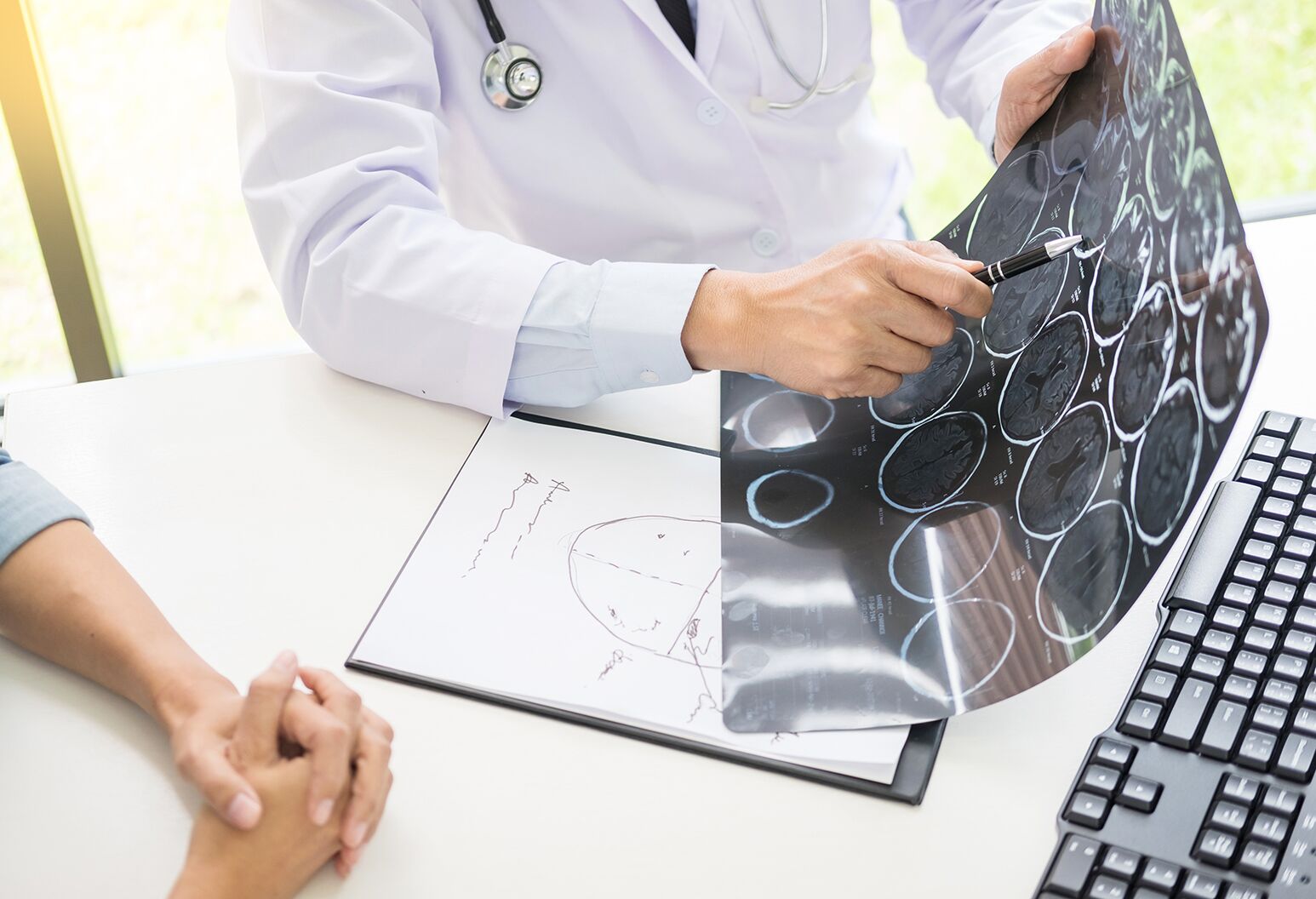
Doctors told Espinoza she had vertigo, but that didn’t explain her tingling fingers. She followed up with her primary care physician, and then a specialist, but had a hard time believing she had a simple case of vertigo. Her instincts were right. An MRI revealed something called a venous anomaly — an abnormal formation of blood vessels in her brain — and it had led to a brain bleed.
“I came home and let my mom know,” Espinoza says. “Everyone was very worried. I was like, 'This is not me. This can’t be me.'”
The doctor told her the bleed should stop on its own; they could just monitor it. That didn’t reassure her — what if it happened again? So, once again, she sought a second opinion. Soon, she was sitting with neurosurgeon Thomas Link, MD, at North Shore University Hospital (NSUH).
Reviewing her MRI, Dr. Link finally gave Espinoza a diagnosis. She had a cerebral cavernous malformation (CCM), also called a cavernoma.
A cavernoma is a type of angioma, or a tightly packed cluster of small, abnormally shaped blood vessels. What distinguishes cavernomas from other angiomas is their location in any of several areas of the nervous system. Cavernomas — also known as cavernous malformations, cavernous angiomas, intracranial vascular malformations or cavernous hemangiomas — look something like a berry and can be as small as a quarter-inch or as large as 4 inches.
Cavernomas are surprisingly common and occur in about 1 in 100 to 200 people.
At least 30 percent of patients living with a cavernoma experience some symptoms — most are in their 20s or 30s. Another 25% experience no symptoms at all. Symptoms can be related to the part of the brain affected by the condition and may appear or subside as the cavernoma changes in size due to bleeding and reabsorption of blood.
Cavernomas that leak blood into surrounding brain tissue, like Espinoza's, can result in lasting neurological problems like:
The only way to prevent a brain bleed from a cavernoma is to surgically remove it. Unfortunately, Espinoza’s cavernoma was in her brain stem, which regulates many of the body’s most basic functions: breathing, heart rate, blood pressure and more. Operating would be so risky that most neurosurgeons would not even attempt it, Dr. Link told her; instead, the typical recommendation for a problem like hers — a brainstem cavernoma — was to simply watch it, unless multiple brain bleeds left no other choice but to accept the risks of surgery.
Espinoza left the office feeling defeated, and unsure of her next steps. But a few days later, Dr. Link followed up with a suggestion: Espinoza should make an appointment with his colleague, Amir Dehdashti, MD, director of cerebrovascular neurosurgery research at NSUH. Dr. Dehdashti specialized in problems like hers, Dr. Link told Espinoza, and was one of just a few neurosurgeons in the country with extensive experience in removing brainstem cavernomas.
“He said, ‘I discussed your case with him and we think there may actually be a reasonably safe approach to your particular cavernoma,’” Espinoza remembers. “‘I would like you to see him and hear him out.’”
A renowned neurosurgeon and one of the few dual-trained cerebrovascular and skull base surgeons in the United States, Dr. Dehdashti treats more patients with this condition than any surgeon in the region. His manner was reassuring as he told Espinoza he thought she could be treated.
“Cavernous malformations that are deep in the brain, like hers, present a much higher risk of bleeding,” says Dr. Dehdashti. Espinoza’s imaging tests showed that she’d already experienced more than one small hemorrhage; the next one could be much more dangerous. Her cavernoma was dangerously close to brain regions responsible for her facial muscles, eye movement, hearing and physical coordination — a bleed could impact all of those or even leave her in a coma.
“The constant anxiety, not to mention the potential she faced for future damage, would greatly compromise her quality of life,” he says.
Surgery would be difficult, Dr. Dehdashti acknowledged. But he felt sure he could find a safe pathway to removing the cavernoma.
Espinoza knew she was facing a life-changing decision, but she felt no doubt. “I knew from the second he started speaking about it that I wanted to go through with the surgery,” she says. “If I left it alone, my risk of having another brain bleed would go up every year. I wanted my life back.”
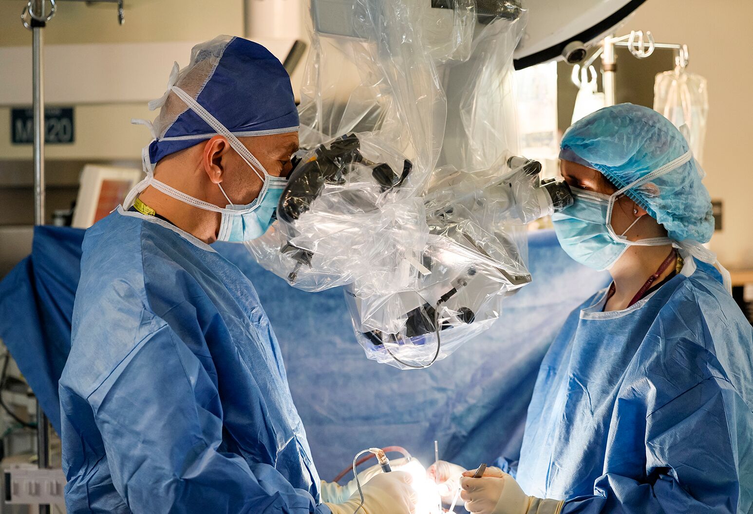
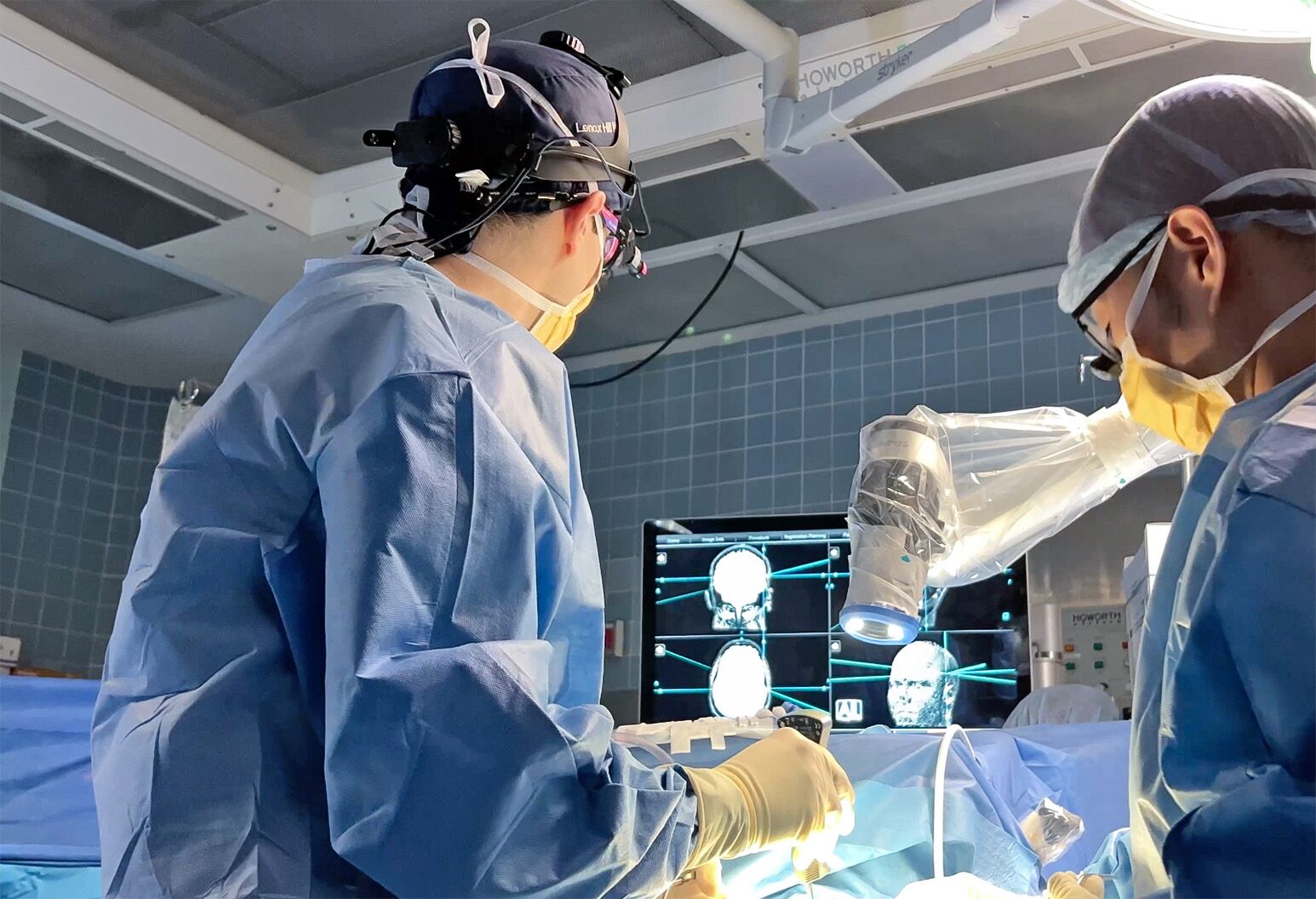
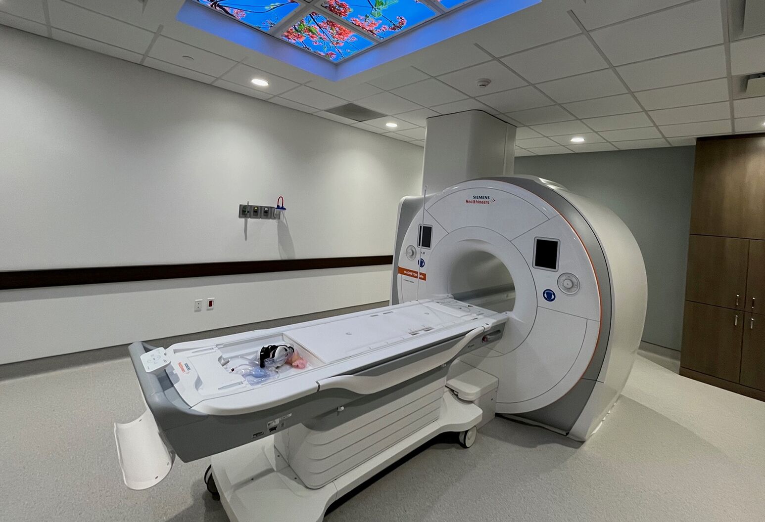
Espinoza's surgery, which took place on June 23, 2022 at NSUH, took five hours and involved state-of-the-art tools. Guided by intraoperative brain-stem mapping, the surgical team used a probe to electrically stimulate nerves in order to identify the areas that controlled vision, speech and movement. This process helped them determine a safe route to the cavernoma.
In addition, Espinoza’s brain and cavernoma had been imaged in detail using a specialized CT scanner. The scans were uploaded into a workstation in the operating room, precisely lined up with bony landmarks of her skull, and projected in 3D onto her brain to help the surgical team locate the exact contours of the abnormal blood vessels.
Carefully, Dr. Dehdashti navigated under the surface of the brain stem to find the cavernoma. Then he dissected around it and took it out. It was almost an inch long — almost half the width of the brainstem in that spot.
When Espinoza woke from surgery, she asked about her cats — were they okay? “That was a great sign,” says Dr. Dehdashti. “She was speaking without any problem and showing no signs of damage.”
Still, she needed time to recover. She had double vision and her right side was weak and numb; her arm and leg didn’t work quite normally. This degree of temporary dysfunction was to be expected after such a delicate surgery. But within a day or two, she was walking the hallways of the hospital. On day three, she went home and within a week she was walking on her own.
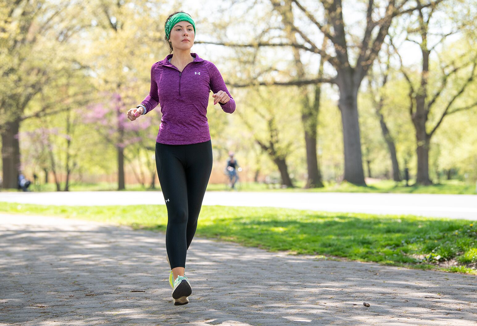
Physical therapy helped a lot. She did exercises to strengthen her right hand and leg and improve her balance, and then she started on the treadmill. Short walks became longer walks and then short runs. In March, she ran her first post-surgery 10K race.
The best news of all is that follow-up MRI scans showed that the cavernoma is completely gone. “Her brain looks perfect,” Dr. Dehdashti says.
Espinoza is ready for the next phase of her life: She and her boyfriend, Garrett Burns, 33, recently moved to a new apartment in Brooklyn, and she is planning to run a marathon next year. They feel lucky that Espinoza trusted her gut when she felt she had more than just vertigo — and that she found her way to a surgeon with the highly specialized skills needed to treat her particularly challenging problem. Espinoza has joined a cavernoma advocacy group and is passionate about sharing information with other patients.
“I’m just thankful that I had access to a very, very good neurosurgeon who was able to give me my life back,” she says. “I feel like I’m born again.”
Our representatives are available to schedule your appointment Monday through Friday from 9am to 5pm.
For a Northwell ambulance, call
(833) 259-2367.