Our representatives are available to schedule your appointment Monday through Friday from 9am to 5pm.
For a Northwell ambulance, call
(833) 259-2367.
People like to compare the brain to a sponge, soaking up knowledge and experiences as we grow. Also like a sponge, the brain can swell with fluid, a much more serious event: This can happen after an injury to the head and the swelling can cause the brain to press up against the skull. In severe situations, surgeons may have to remove part of the skull to relieve the pressure.
While this allows the brain to heal, the missing piece of skull leaves the brain vulnerable to blows and can impair its function. Cranioplasty is a surgery that can make the skull whole again either with a bone graft or a cranial implant made of synthetic materials like medical-grade plastic or metal. Today, these implants are 3D printed, improving the fit and reducing complication rates.
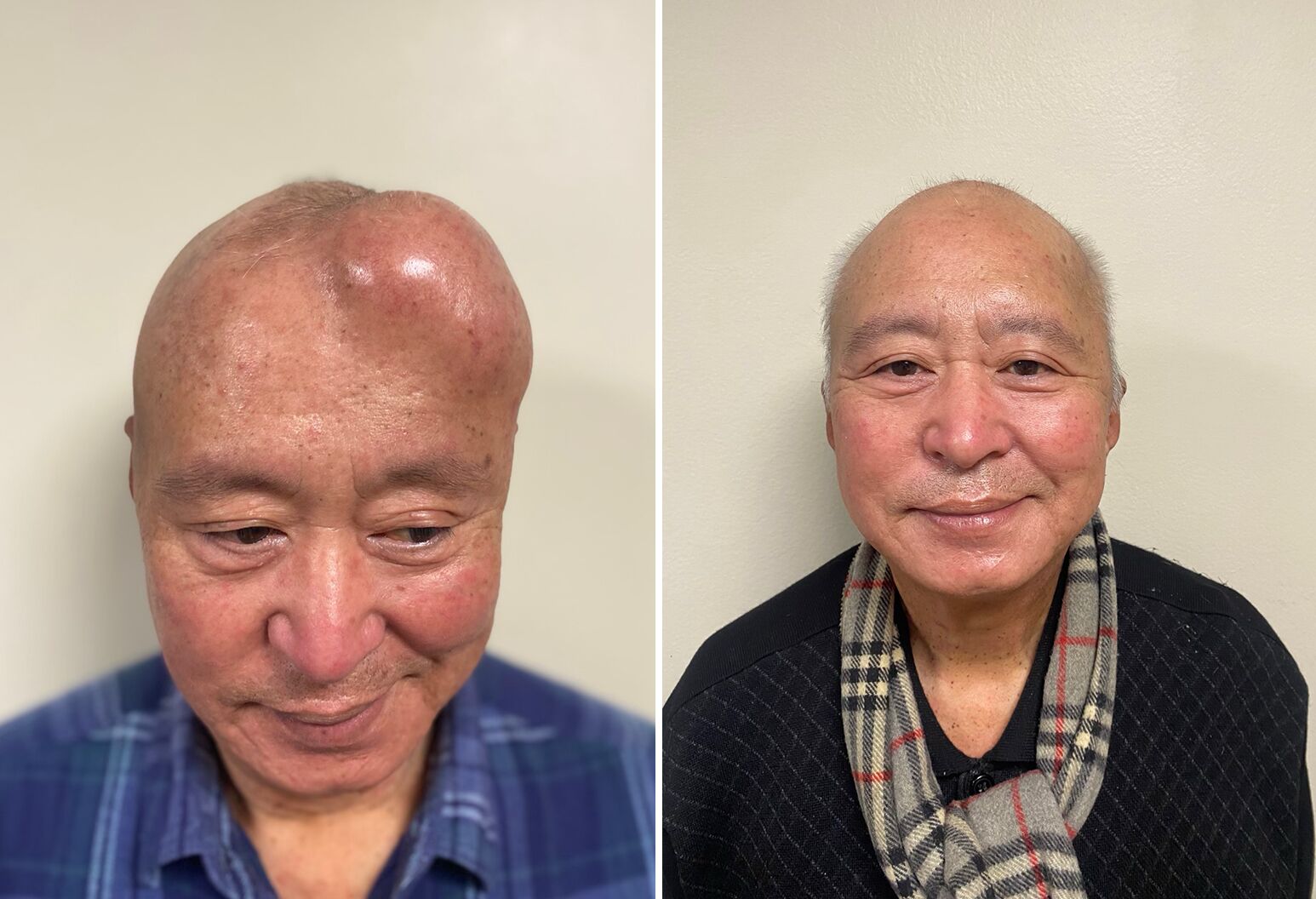
Cranioplasty can rapidly resolve neurological symptoms that come with missing a part of the skull. It also restores a person’s appearance and sense of self. Prior to cranioplasty, the skull may appear sunken or full in on one side, explains Netanel Ben-Shalom, MD, a neuroplastic and reconstructive surgeon at Lenox Hill Hospital.
“The deforming asymmetry can be striking — each time a patient looks in the mirror, it brings them back to the diagnosis, to the fear, to the trauma,” says the world-renowned expert, who leads Lenox Hill’s neuroplastic surgery program, the first of its kind in New York State. “My goal with cranioplasty is to reconstruct that so patients regain their confidence and sense of self next time they look in the mirror.”
If you or a loved one is considering cranioplasty, this guide will help you understand the procedure, the different materials used, and what to expect during surgery and recovery.
Cranioplasty is a surgical procedure designed to repair defects in the skull. It blends neurosurgery with plastic and reconstructive techniques. The surgery can last between two to four hours, depending on the size and complexity of the skull deformation. Trauma to the head and the surgery to remove part of the skull (craniotomy or craniectomy) are two common reasons one would need cranioplasty. Others include:
When left exposed, the brain is vulnerable to direct injury and doctors may advise patients to wear a helmet when moving around. Additionally, some people experience symptoms related to the loss of bone. “The brain doesn’t function the same when it’s not protected with the bone,” Dr. Ben-Shalom explains. Patients often deal with:
These symptoms can vary from mild to severe and happen because of changes to the pressure inside the head. “Normally, the skull provides a protective barrier that keeps everything balanced,” he says. ”When the brain is only covered by skin and soft tissue, outside pressure pushes in on the brain, altering your normal appearance and the fluid dynamics that keep everything working normally.”
The added pressure can also disrupt blood flow, reducing the amount of oxygen certain areas of the brain receive. “The cortical blood vessels actually constrict because of this added pressure, resulting in reduced cerebral blood flow to critical regions,” Dr. Ben-Shalom says. This compressive effect also changes the flow of cerebrospinal fluid (CSF), which delivers nutrients and acts as a cushion for the brain. The result is impaired functions like balance, memory and even mobility. Cranioplasty helps restore normal brain function by rebuilding that protective barrier.
“We see patients who cannot move half of their body before cranioplasty,” Dr. Ben-Shalom notes. “After surgery, patients often regain lost function — and quickly. I remember a patient who could not talk for four months and was very sleepy. Just two days after the cranioplasty she started to speak.”
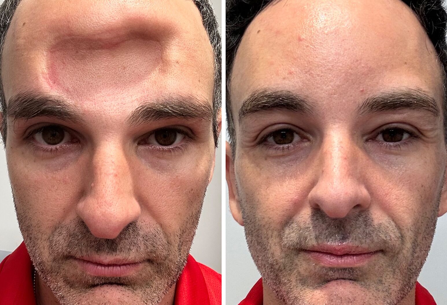
Ido Simyoni, marathon runner, was left with a mass on his forehead due to a condition called fibrous dysplasia, in which bone is replaced with scar tissue. Lenox Hill neurosurgeons performed Simyoni's cranioplasty in 2023. Photo credit: Northwell Health.
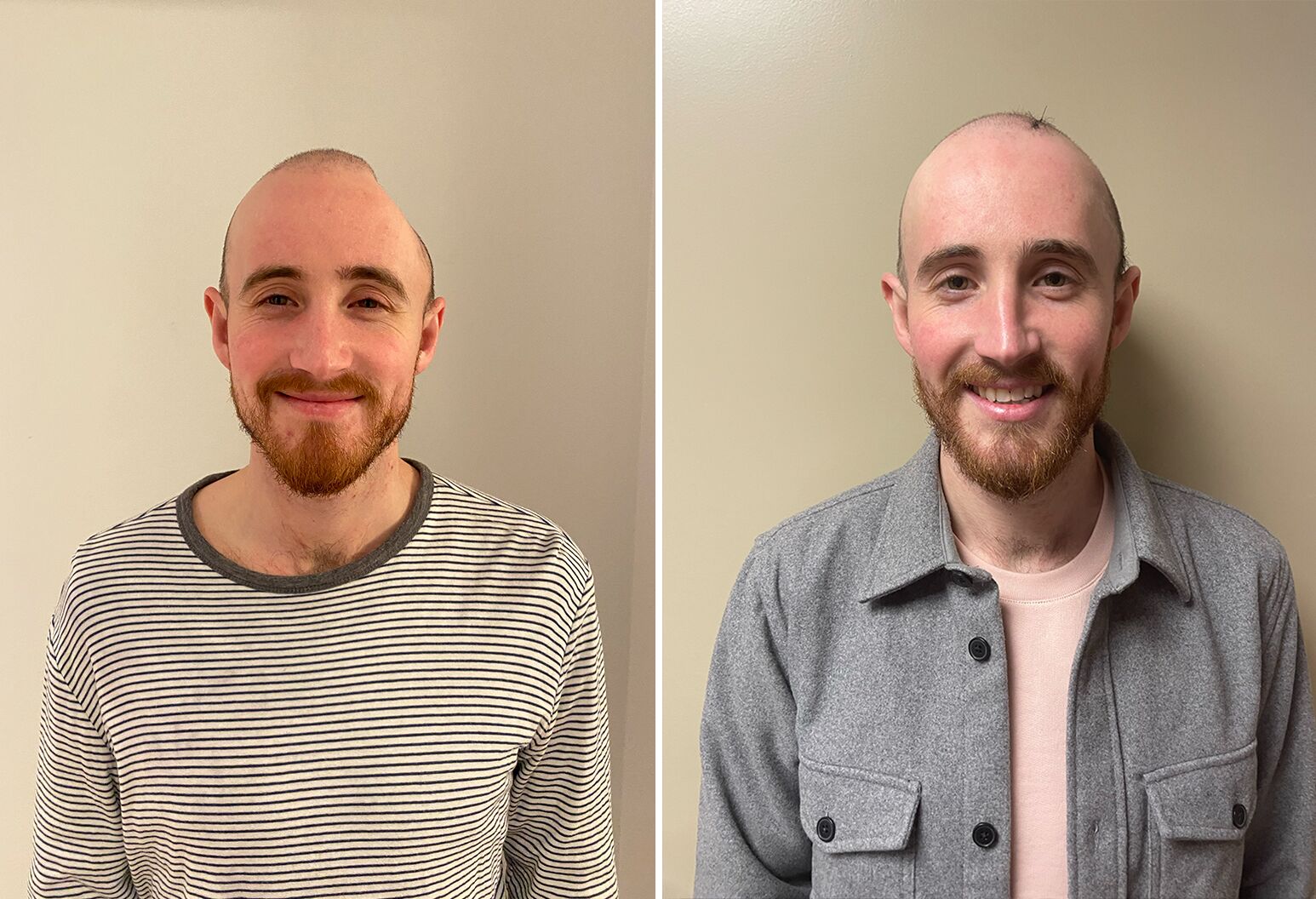
Tucker Marr had cranioplasty surgery at Lenox Hill Hospital in February 2022 to repair damage to his skull after a traumatic brain injury. Photo credit: Northwell Health.
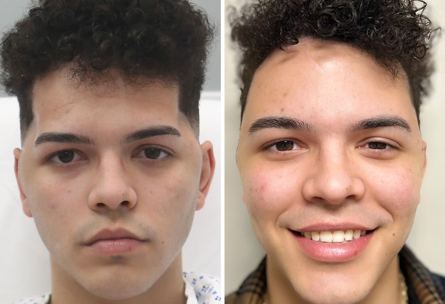
To help Anthony Mercado survive a serious car crash, SIUH neurosurgeons had to remove a portion of his skull to relieve brain swelling, leaving him with skull deformities. Dr. Ben-Shalom performed a cranioplasty, restoring skull appearance and facial muscle function. Photo credit: Northwell Health.
Previously diagnosed liver cancer turned into a mass on the left side of Heju Li's brain. In one procedure, Dr. Ben-Shalom and his team removed the tumor and reconstructed the portion of his skull that was being invaded by the tumor. Photo credit: Northwell Health.
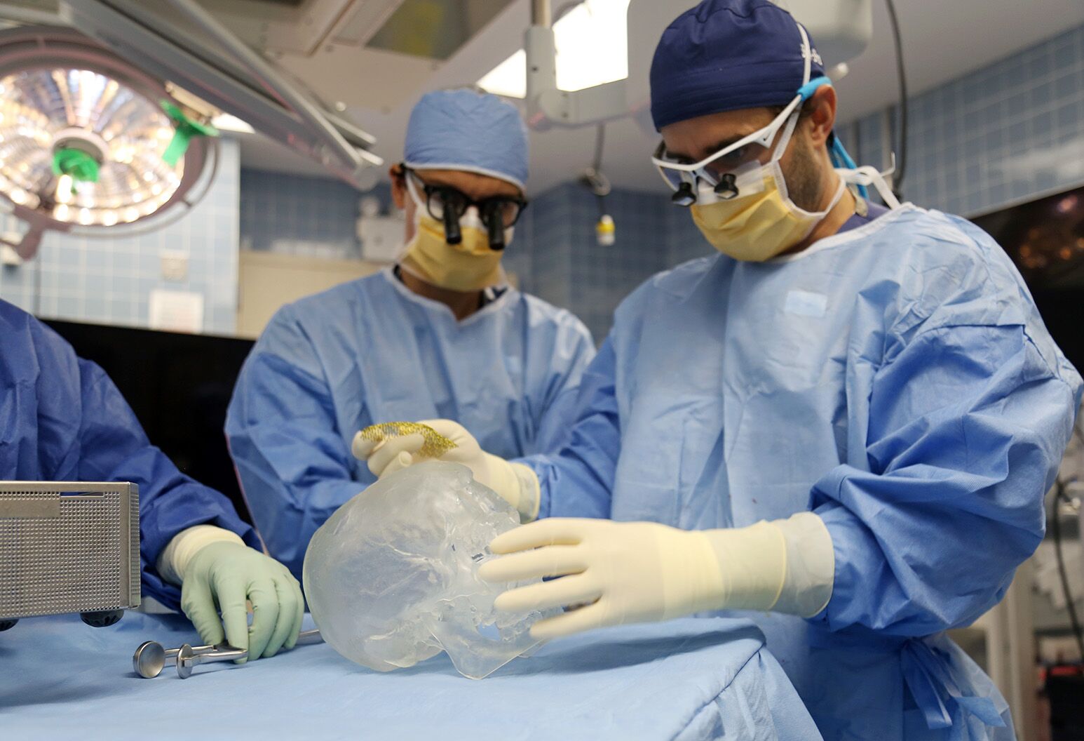
To replace the missing bone, surgeons may use a patient’s own bone (known as an autologous transplant) or synthetic materials such as acrylics mesh or medical-grade titanium. When paired with advanced imaging and 3D modeling, surgical teams using synthetic materials can tailor the implant to the exact measurements of the patient’s skull defect. This approach, called virtual surgical planning, ensures that the implant integrates well with the patient’s tissue, reducing the likelihood of complications related to a bad fit or movement of the implant over time.
Dr. Ben-Shalom says this has been a major advance that has helped to reduce complications and improve how patients look and feel. “We take everything into consideration: the location of the defect, the type of implant, the patient’s age, medical history and even their lifestyle and goals post-surgery,” he says.
Dedicated neuroplastic teams like the one at Lenox Hill Hospital are also making a significant difference. Unlike general neurosurgical teams, a neuroplastic team specializes in both the functional and cosmetic aspects of skull reconstruction. “While a neurosurgeon is focused solely on the engine of the car, the neuroplastic surgeon focuses on both the engine and the hood,” Dr. Ben-Shalom says.
In addition to training as a neurosurgeon, neuroplastic surgeons complete additional study in plastic and reconstructive surgery. This cross-section of skills remains unique, with just a handful of dedicated neuroplastic programs in the country. “And it allows us to focus on every detail: from scalp rearrangement to skull base reconstruction. We are laser-focused on preserving our patients' form and function, tailoring our approach to their unique needs,” Dr. Ben-Shalom explains.
Choosing what material to use for cranioplasty is complex, he explains, and requires weighing the strengths and weaknesses of each.
There are several types of medical-grade plastics used for cranioplasty surgery — these are also used to construct dental implants, crowns and caps. Two common types are PEEK and PMMA.
Polyetheretherketone (PEEK) is highly compatible with human tissues, reducing the risk of immune rejection and inflammation. It is also comparable to bone in its strength and elasticity, making it a very durable implant. PEEK is also the most expensive material used in reconstructive surgery.
Polymethyl methacrylate (PMMA) is the material used to produce acrylic implants, often referred to as PMMA cranioplasty, or “bone cement.” It is strong but lacks the elasticity of PEEK and is more prone to cracking under extreme stress. PMMA is highly biocompatible and relatively inexpensive compared to other materials.
Clear PEEK and PMMA are both also radiolucent, meaning they can transmit ultrasound waves. “When patients come for follow-ups, I can take an ultrasound probe and look at their brain,” Dr. Ben-Shalom says. “We can confirm everything looks normal and detect any problems right there, non-invasively.”
Titanium mesh is a lightweight, durable metal commonly used for cranioplasty. It is strong and less likely to corrode or degrade over time. This is crucial in surgical implants, which are intended to last for many years. It integrates well with the body’s tissues, reducing the risk of rejection or adverse reactions. Like any material, improper positioning and technique can cause discomfort or pain, as well as higher risk for complications.
While titanium is generally well-tolerated, sensitivity or allergic reactions may occur in rare cases.

This procedure involves reattaching a patient's own bone, which was removed during surgery, to repair a defect in the skull. This approach significantly reduces the risk of rejection, as the body recognizes its own tissue, promoting better integration with surrounding structures.
Using the patient’s own bone isn’t always an option if it becomes infected after removal. Even when preserved perfectly, Dr. Ben-Shalom explains, there is a high risk of infection and resorption (where the bone deteriorates over time), compared to synthetic materials. This can compromise the repair.
Another issue: Once a portion of bone is removed from the skull, it loses its blood supply.
Dr. Ben-Shalom’s team is now involved in developing what he calls next generation implants. “Not only are they replacing the hard coverage of the brain, but also the soft tissue.” These soft tissues include muscle, fat, connective tissue and even blood vessels.
“Now, our implants can compensate for soft tissue defects. These implants have the potential to house technologies, which will allow us to retrieve biometric data from the brain.”
These advancements in surgical techniques and implant materials are allowing more patients to undergo cranioplasty with fewer issues and a quicker return to normal life.

Most cranioplasty patients spend several days in the hospital for monitoring. Medical teams watch for any complications like bleedings and infections, which are high, compared with other types of brain surgery.
Historically, cranioplasty complications have included issues such as infection, poor healing of the surgical site and the body rejecting the materials used for the implant. Studies show that between 35% and 40% of patients experience complications, with infection being the most common issue.
But that is starting to change, thanks to new technology and dedicated teams specializing in this type of reconstruction, Dr. Ben-Shalom says. “I see many patients that come in with failed reconstructions due to infection, rejection or multiple surgical attempts. Patients that have exhausted all traditional treatment options come to Lenox Hill Hospital to meet our team. We offer a personalized approach, even in the most complex cases. When others see challenge, we see opportunity.
"We are committed to delivering the most innovative combination of surgery and science to each patient."
Following cranioplasty surgery, patients often report feeling better both physically and mentally. Dr. Ben-Shalom explains that a patient’s outlook and emotional state can influence recovery: “Patients who are happier and more confident tend to have better outcomes overall. This underscores the importance of not just restoring the skull’s structure but also helping patients regain their confidence and sense of well-being.”
For a smooth recovery, Dr. Ben-Shalom recommends the following steps to reduce the risk of complications.

Our representatives are available to schedule your appointment Monday through Friday from 9am to 5pm.
For a Northwell ambulance, call
(833) 259-2367.