Our representatives are available to schedule your appointment Monday through Friday from 9am to 5pm.
For a Northwell ambulance, call
(833) 259-2367.

Cataracts and glaucoma are two eye conditions that can rob your vision, and both are quite common. More than 24 million Americans will experience cataracts at some point, and three million will develop glaucoma. These diseases are somewhat similar, but they are diagnosed and treated differently.
“The main difference is that surgery for cataracts can restore vision, whereas the damage caused by glaucoma is permanent,” says Victoria Rohring, DO, MS, an ophthalmologist with Northwell Health’s Eye Institute. “The good news is that most preventive eye health measures work for both glaucoma and cataracts. What prevents one often will prevent the other.”
Here’s what you need to know about glaucoma and cataracts.
Glaucoma is a buildup of pressure in the eye that can damage the optic nerve. Left untreated, it can eventually cause blindness. About 1.5 million Americans living with glaucoma are undiagnosed. While it’s most frequently diagnosed in older adults, it can occur at any age.
Tiny channels that run between the cornea and the iris drain excess eye fluid. These channels can become obstructed, and as fluid builds up in the eye, intraocular pressure (or IOP) increases, leading to glaucoma. “It's like tire pressure,” Dr. Rohring explains. “The pressure has to be just right. Too much or too little can cause problems.”
Given time, this pressure can damage the optic nerve. Common risk factors for glaucoma include:
There are several different types of glaucoma, and they can vary in cause and severity:
Part of what makes glaucoma so troublesome is that it is often asymptomatic in its earlier stages, says Dr. Rohring. “By the time patients notice something is wrong, they’ve usually lost much of their peripheral vision. But you don’t lose your eyesight all at once. People with glaucoma typically retain their central vision until the later stages of the disease. We call this tunnel vision, which can make it hard for patients to function because their depth perception is off, and things may appear dimmer at times.
“This is what prompts most patients to come in and that's when we find they're already in the late stages of the glaucoma.”
There is one type, acute angle-closure glaucoma, that can cause symptoms before vision loss. Those include excruciating eye pain, nausea and sensitivity to light. Generally, though, a patient won’t have the luxury of such warning signs, Dr. Rohring explains, which is why regular testing is paramount for people of all ages.
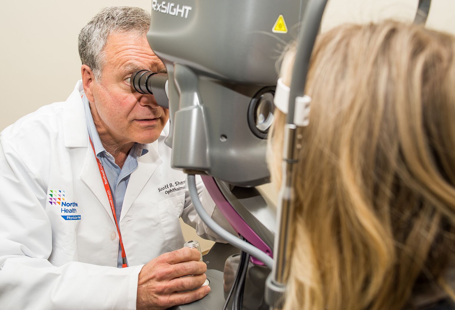
"Early detection is crucial for managing glaucoma effectively,” she says. “Diagnosing the condition involves a thorough examination of the eye, particularly the optic nerve, which is where the damage occurs.”

There are several tests ophthalmologists use to help them determine the presence and/or severity of glaucoma.
Tonometry measures the intraocular pressure inside the eye. The most common method is applanation tonometry, which gently applies pressure to the cornea. “We apply a little bit of pressure to the outside of the eye, and then we see how much rebound pressure we get back,” Dr. Rohring says.
Another technique called pneumatic tonometry, also known as the “air puff” exam, uses a quick pulse of air to slightly depress the cornea, recording the degree of deformation. “This isn’t used as often because it has shown to be less accurate,” she says.
Gonioscopy involves the use of a goniolens (a special type of contact lens), which is held against the eye to observe the angle between the iris and cornea. Consequently, this test is helpful in diagnosing narrow- or closed-angle glaucoma. “This helps us evaluate if fluid is draining properly, or if it's partially or completely obstructed,” Dr. Rohring says.
Fundus photography provides imaging of the fundus — a structure at the back of the eye that houses the optic nerve. A compromised or weakened optic nerve could be a sign of glaucoma.
Optical coherence tomography (OCT-RNFL) uses light waves to take a more detailed cross-sectional image of the optic nerve, helping to measure its thickness. OCT-RNFL is used to monitor the loss of nerve fibers in glaucoma patients over time and assess the progression of the disease.
Visual field test assesses peripheral vision loss, a key indicator of glaucoma. The patient is asked to detect lights shown in their peripheral vision, and any blind spots are noted.
Pachymetry measures the thickness of the cornea, as thinner corneas can increase the risk of glaucoma.
By taking the right steps, you can lower your risk of losing sight to glaucoma. "Regular exercise and a healthy diet can go a long way in keeping glaucoma at bay,” Dr. Rohring says. “When possible, avoiding excessive consumption of steroid medications and caffeine can also help keep eye pressure low.”
Annual eye exams are essential to detect early signs of eye disease, even before symptoms appear. They are recommended for patients 55 and older, but those with risk factors like family history, diabetes, or trauma to the eyes should start younger. Adults younger than 55 with no risk factors should have a comprehensive eye exam every two years.
Despite a patient’s best efforts, however, prevention can prove unsuccessful. When it does, treatment will be necessary.
You’ll be pleased to learn that there are a number of effective treatments for glaucoma. “All of our treatment is based on controlling the pressure and we typically start with the non-invasive options first,” Dr. Rohring says.
Glaucoma treatment, in most cases, begins with eye drops. The mechanism of the eyedrops’ action varies. Some eye drops work by increasing fluid drainage in the eye. Those include:
Others reduce the amount of fluid that the eye produces, in turn lowering the burden on the drainage channels. Those include:
Still others like alpha agonists combine both mechanisms, making them convenient for patients who need multiple medications.
“Eye drops are only effective if patients adhere to a strict schedule,” Dr. Rohring says. “Some experience side effects like eye irritation or redness, but these can usually be managed by adjusting the treatment regimen.”
Laser procedures are increasingly used to manage glaucoma, especially when eye drops aren’t effective or cause side effects. These minimally invasive options have fewer side effects and shorter recovery times than traditional surgery. The two most common types of glaucoma laser procedures are:
Selective laser trabeculoplasty (SLT): A short laser procedure used to treat open-angle glaucoma by lowering eye pressure. SLT targets specific cells in the drainage angle of the eye, stimulating a biological change that improves fluid drainage. While effective — about 75-80% of patients see a significant reduction in IOP — this is not a permanent solution and the procedure may need to be repeated. Results typically last for 1-5 years.
Laser peripheral iridotomy (LPI): Indicated for narrow-angle glaucoma, a condition where the iris physically obstructs the drainage passageway. This laser surgery creates a small opening in the iris, allowing fluid to bypass the obstruction and flow freely, relieving pressure. With a high success rate, LPI typically provides long-term relief.
When glaucoma doesn’t respond to the previous options, more invasive surgical options are available. Two procedures make up the bulk of traditional glaucoma surgeries:
“Trabeculectomy is generally considered more effective, while tube shunt surgery is less likely to produce complications,” Dr. Rohring says.
Whether eyedrops, laser or traditional surgery, all glaucoma treatments are considered “secondary prevention” measures — meaning they don’t reverse damage to the optic nerve that has already occurred. Rather, they aim to prevent further progression of glaucoma and preserve as much nerve function as possible.
This eye condition is a clouding of the usually clear lens, and it blurs your sight. “It’s like looking through a dirty glass window,” Dr. Rohring says. “For the light to get inside the eye it must pass through the lens and if the lens is cloudy then you're not going to be able to see well.”
As is the case with glaucoma, untreated cataracts can cause partial or even total vision loss; they’re responsible for more than half of all cases of blindness worldwide.
The eye’s lens is composed of proteins. With age, these proteins can break down, clump together and form clusters. The result is a cataract, which clouds the lens and blocks light from reaching the retina.
Risk factors for the development of cataracts include:
“If you live long enough, you will likely develop some form of cataracts,” Dr. Rohring says.
There are three main types of cataracts, distinguished by their point of origin on the lens.
Fortunately, it’s possible to spot cataracts early. Unlike glaucoma, cataracts produce symptoms, plus they’re actually visible — the protein clumps produce a milky hue in the lens.
People with the disease experience varying degrees of blindness depending on how far the cataracts have progressed.
In the early stages of cataracts, the clouding of the lens is minimal. Vision begins to blur slightly, particularly in areas of fine detail, but it may be hard to notice at first. People may have trouble reading small text or recognizing faces at a distance.
As the cataract develops, the vision becomes more blurred and less sharp, especially in lower lighting conditions. This stage is often characterized by a gradual increase in glare or halos around lights, making it difficult to drive at night. Colors may begin to appear less vibrant, and the lens starts to take on a dull or faded appearance.
By the time the cataract reaches the mature stage, the lens has become significantly clouded, making it hard to see clearly in low-light or nighttime conditions. Daytime vision may also be noticeably reduced, as the cloudiness in the lens blocks more light, causing the vision to appear hazy and foggy.
In the final stage, the cataract becomes completely opaque, and the lens appears yellowed or even milky. At this point, the vision loss is significant, and the patient is likely to be nearly or completely blind. The lens may also harden, making it difficult for the surgeon to remove it during surgery. Additionally, the pressure inside the eye may increase, potentially leading to secondary glaucoma, a condition where the increased pressure further damages the optic nerve and worsens vision. In this stage, cataract surgery becomes necessary to restore vision.
As is the case with glaucoma, cataracts can be slowed or prevented by following healthy habits. Such measures include:
If your vision becomes so impaired that you’re struggling to read, drive or watch television, your doctor may recommend cataract surgery. Currently, there aren’t any non-surgical treatments for cataracts.
Cataract surgery is typically an outpatient surgery and eye surgeons can do the procedure with local anesthesia. A patient's clouded lens is removed, and an artificial lens is inserted in its place. Unlike glaucoma treatments, surgery for cataracts can restore vision. “It depends on how cloudy their lens is, but patients can go from just seeing shadows to regaining 20-20 vision after cataract surgery,” Dr. Rohring says. “The last couple of decades have brought tremendous improvement in our surgical technique, patient outcomes and safety.”
Recent developments in cataract surgery include:
To find out more about chronic eye diseases, click here.
Our representatives are available to schedule your appointment Monday through Friday from 9am to 5pm.
For a Northwell ambulance, call
(833) 259-2367.